


In situ study of phase transition in HZO ferroelectric thin films via TEM electron beam irradiation

 ,
, Abstract
Oxygen vacancies (VO) play a crucial role in the stability of the ferroelectric orthorhombic (o-) phase of hafnium dioxide (HfO2)-based thin films. However, the stability of the ferroelectric phase of HfO2 under the action of VO and the mechanism of ferroelectric phase transition are still unclear. In this work, VO concentration in Hf0.5Zr0.5O2 (HZO) thin films is tuned through electron beam irradiation inside a transmission electron microscope. For the crystalline HZO thin films processed through rapid thermal annealing, the increase of the VO concentration during in situ electron beam irradiation facilitated the phase transition from the non-polar monoclinic (m-) and tetragonal (t-) phases to the polar o-phase. For the amorphous HZO thin films, the nucleation and growth process of the m- and o-phases are observed during in situ electron beam irradiation. The phase transition from m-phase to o-phase is accompanied by the evolution from tensile to compressive strain. These results help to clarify the mechanism of ferroelectric phase transition under the action of VO, and guide the control of the ferroelectric properties and phase stability of HfO2-based thin films.
Keywords
INTRODUCTION
Ferroelectric materials have great potential in electronic information technology, especially in ferroelectric storage and computing devices, such as ferroelectric field-effect transistors and nonvolatile memories[1,2]. However, traditional perovskite-structured ferroelectric thin film materials suffer from poor compatibility with complementary metal-oxide-semiconductor (CMOS) technology, limited miniaturization, low storage density, and environmental issues such as lead pollution[3]. In contrast, hafnium dioxide (HfO2)-based ferroelectrics offer a promising alternative due to their excellent CMOS compatibility, wide bandgap, high storage density, low energy consumption, and scalability to nanoscale dimensions[4,5]. The ferroelectricity of HfO2 was first discovered in 2011 in Si-doped HfO2 thin films[6], a breakthrough that has since spurred widespread interest in both ferroelectric materials and memory devices[7]. Subsequent research has shown that the ferroelectricity of HfO2-based thin films originates from the non-centrosymmetric Pca21 ferroelectric o-phase[8]. Recent studies have further demonstrated the immense potential of HfO2-based systems: for instance, atomic-scale ferroelectric tunnel junctions with unprecedented scaling have been reported[9], and record-high 300 K-resistance switching in ferroelectric-gated Mott transistors has been achieved[10]. However, multiphase, including orthorhombic, monoclinic (m-), and tetragonal (t-) phases, coexist in the atomic layer deposition (ALD)-fabricated HfO2-based thin films due to the metastable characteristic of the o-phase[11-13]. Therefore, controlling and stabilizing the ferroelectric o-phase is critical to achieving superior device performance of HfO2-based thin films[14].
Oxygen vacancies (VO) are an important factor affecting the stability of the HfO2 o-phase; thus, the ferroelectricity of HfO2 can be regulated by tuning the VO[15,16]. First-principles density functional theory (DFT) calculations confirmed that increasing the concentration of VO within a certain range is beneficial to the stability of the o-phase because the presence of VO promotes the generation of the o-phase[17]. Furthermore, VO in HfO2 can exist in several charge states. Most first-principles studies typically consider at least three major charge states: the neutral vacancy (VO0), the singly positively charged vacancy (VO+), and the doubly positively charged vacancy (VO2+). In many cases - especially in the context of ferroelectric phase stabilization - the doubly charged state (VO2+) has been highlighted as particularly significant because its formation and lower diffusion barrier help to promote the phase transition from the non-polar to the ferroelectric state[16,18]. VO can be introduced into HfO2 by optimizing deposition conditions such as O reactant species and deposition temperature, and doping of bi- or tri-valent chemical species to meet the charge neutrality condition, etc.[19-21]. The concentration of VO in ALD-fabricated HfO2-based ferroelectric thin films could be reduced by oxygen plasma (O2 Plasma) treatment or O3 treatment, macroscopically manifested as decay of ferroelectric performance[22]. The decrease in VO concentration changes the stability of each phase in the film, which reduces the content of the o-phase evidenced by phase composition characterization experiments[15,23]. It was found that in situ irradiation of Hf0.5Zr0.5O2 (HZO) ferroelectric thin films with an electron beam can induce the generation and redistribution of VO, causing local stress fields, thereby affecting the stability and migration of 90° ferroelectric domain walls[24]. At the same time, the local stress field caused by the generation and redistribution of VO plays an important role in the ferroelectric phase transition. Up to now, the results of theoretical calculations and experiments of VO or strain effects on the stability of o-phase are not consistent. In DFT calculations, it is believed that in-plane compressive stress is beneficial for the stability of the o-phase, while in-plane tensile stress significantly promotes the stability of the m-phase[25-27]. However, in experiments, by applying different stresses to the HfO2-based thin film through different element doping, different thermal expansion coefficient electrode materials, different electrode sizes, and different substrates, it was found that in-plane tensile stress is beneficial for the stability of the o-phase, but not for the stability of the m-phase[6,28,29].
In this work, the high-energy electron beam of a transmission electron microscope (TEM) was used to tune the VO in HZO ferroelectric thin films, and to reveal the effect of VO concentration on the ferroelectric phase transition of HZO thin films and unveil its ferroelectric phase transition mechanism. It was found that the concentration of VO in the HZO ferroelectric thin film significantly increased after electron beam irradiation, accompanied by a phase transition from the m-phase to the o-phase. Meanwhile, geometric phase analysis (GPA) found that during the phase transition of HZO ferroelectric thin films from m-phase to o-phase, the state of strain changes, gradually transitioning from tensile strain to compressive strain. Similarly, we observed the phase transition process from the t-phase to the o-phase, as well as the nucleation and growth of the m-phase and the o-phase under electron beam irradiation in amorphous HZO thin films. These results help to reveal the phase transition mechanism of HfO2-based ferroelectric thin films assisted with VO, guiding the phase stability and ferroelectric performance optimization of HfO2-based ferroelectric thin films.
MATERIALS AND METHODS
HZO ferroelectric capacitors and cross-sectional TEM sample preparation
Fabrication of MFM structure HZO ferroelectric capacitors
For the preparation of the HZO ferroelectric capacitor for ferroelectric performance testing and phase structure characterization, first, a W/TiN bottom electrode with a thickness of about 40 nm was deposited on a heavily doped P-type silicon substrate using magnetron sputtering. Next, with Hf[N(CH3)2]4 (Tetrakis dimethylamido hafnium, TDMAHf) and Zr[N(CH3)2]4 (Tetrakis dimethylamino zirconium, TDMAZr) as precursors and deionized water as the oxygen source, an HZO thin film with a thickness of about 14 nm was deposited on the W/TiN bottom electrode using ALD (Savannah G2 s200, Ultratech/CNTTM) at a deposition chamber temperature of 270 °C and a cycle ratio of Hf precursor to Zr precursor of 1:1. Subsequently, a W/TiN top block electrode or dot electrode with a thickness of about 15 nm was deposited directly or by adding a mask using magnetron sputtering. Finally, the prepared HZO capacitor was rapidly thermally annealed (RTP-150-EP, UniTempTM) at 550 °C for 30 s for crystallization.
HZO ferroelectric film TEM sample preparation
A focused ion beam system (Helios 5 CX, Thermo Fisher ScientificTM) was used to prepare cross-sectional samples of HZO ferroelectric capacitors for characterization by TEM. First, a layer of platinum (Pt) was deposited at the top electrode of the HZO ferroelectric capacitor to protect thin films. Next, a lamella of HZO ferroelectric capacitor sample with dimensions of 10 µm × 2 µm × 8 µm (length × width × height) was cut and lifted out, and welded onto a standard half-copper grid. Finally, the lamella was gradually thinned to a thickness of about 50 nm with decreasing accelerating voltage and Ga ion beam current, thus preparing a cross-sectional sample of HZO ferroelectric thin film for in situ TEM electron beam irradiation.
Structure characterization and ferroelectric performance testing of the fabricated HZO ferroelectric film
The phase composition of the W/HZO/W ferroelectric capacitor was characterized using a grazing incidence X-ray diffractometer (Smartlab 9 kW, RigakuTM). The grazing incidence angle was 0.5°, the scan step was 0.002°, and the scan range was 2θ from 25° to 55°.
The HZO ferroelectric thin film was tested for dynamic hysteresis [polarization-voltage (P-V)] using a ferroelectric analyzer (axiACTT TF analyzer 3000, Germany). P-V curve test conditions: 1 kHz triangular wave, 4 V pulse voltage. Wake-up conditions: 100 Hz square wave, 3.5 V pulse voltage, 1,000 cycles.
HZO ferroelectric film TEM microstructure characterization and in situ electron beam irradiation
A TEM (FEI Talos F200X, Thermo Fisher ScientificTM) was used for TEM characterization of the cross-sectional samples of the HZO ferroelectric thin film and electron beam irradiation with an accelerating voltage of 200 keV. The electron beam spot diameter was scaled down to about 20 nm for continuous irradiation of the HZO thin film. At this time, the electron beam current density was approximately
RESULTS AND DISCUSSION
Characterization of ALD fabricated HZO thin films
The HZO ferroelectric films with Metal-Ferroelectric-Metal (MFM) structures were fabricated through an ALD method as shown in Figure 1A. The specific fabrication process is described in the Section “MATERIALS AND METHODS”. Figure 1B displays the cell structure models of the centrosymmetric

Figure 1. (A) Fabrication process of HZO ferroelectric capacitor, (B) Structural models of a non-polar t-phase with space group P42/nmc and a polar o-phase with space group Pca21. (C) GI-XRD pattern of W/HZO/W ferroelectric thin film, (D) P-V and I-V curves of HZO ferroelectric thin film.
Electron beam-induced VO in HZO films inside TEM
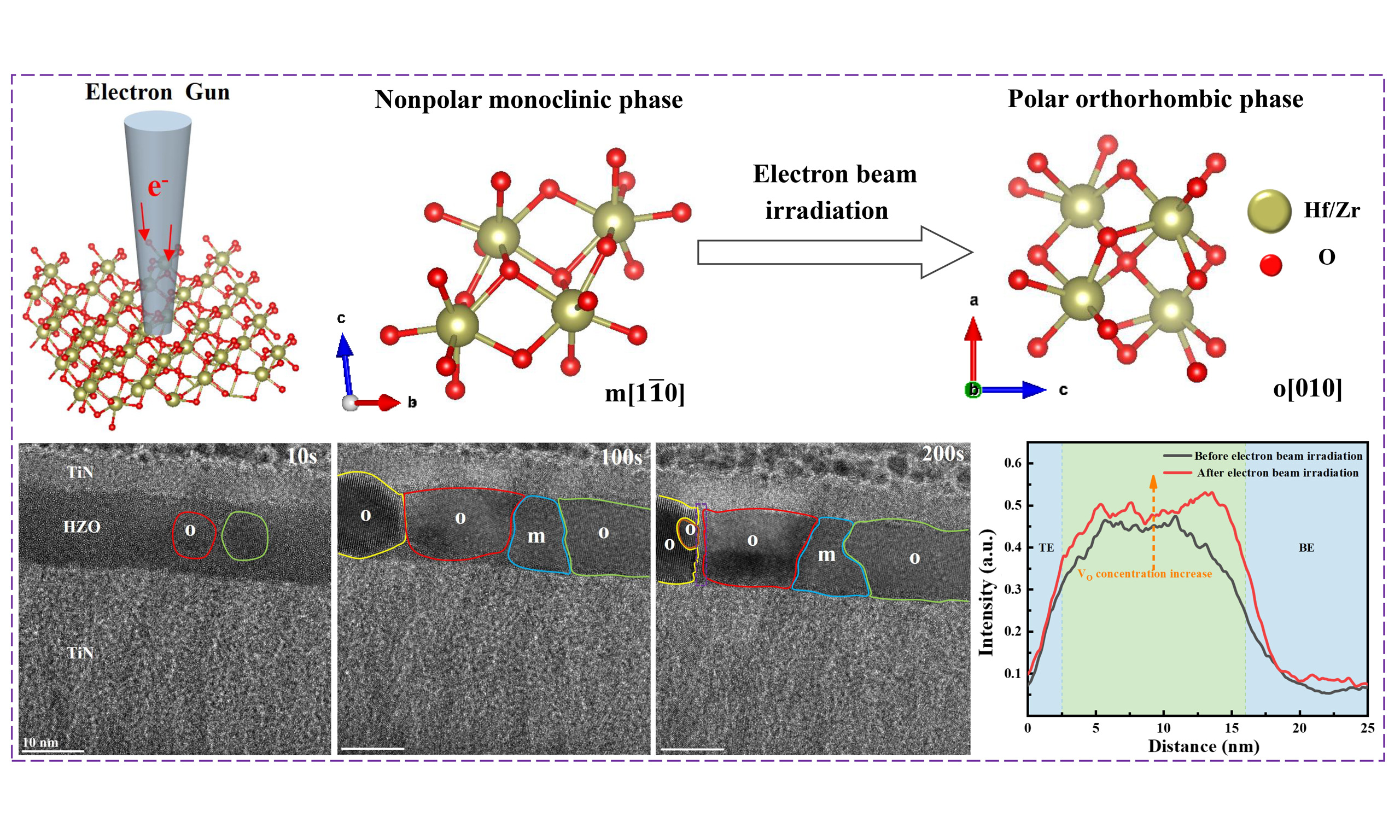
The cross-sectional view of the fabricated HZO film is shown in Figure 2A. The polycrystal HZO layer with a thickness of 14 nm was sandwiched between two W layers (top layer around 15 nm), as shown in the high-resolution TEM (HRTEM) image in Figure 2A. The interfaces between the HZO thin film and the top and bottom electrodes were smooth. In situ electron beam irradiation was carried out inside TEM on the fabricated ferroelectric film as illustrated in Figure 2B, and the VO concentration was analyzed during electron beam irradiation through EDX. By adjusting the diameter of the electron beam, the dosage of the electrons bombarded on the sample area was tuned, and HRTEM images during the irradiation process were captured every minute.

Figure 2. (A) HRTEM image of the cross-sectional morphology of the HZO ferroelectric capacitor, (B) Schematic diagram of electron beam irradiation, scale bar 5 nm, (C) Semi-quantitative analysis of VO concentration before and after electron beam irradiation of the TiN/HZO/TiN ferroelectric thin film, (D) EDX element mapping of the TiN/HZO/TiN ferroelectric thin film before electron beam irradiation, scale bar 10 nm, (E) EDX element mapping of the TiN//HZO/TiN ferroelectric thin film after electron beam irradiation, scale bar 10 nm.
A semi-quantitative analysis of the VO concentration changes in the HZO ferroelectric thin film before and after electron beam irradiation was performed using EDX analysis, with the results presented in Figure 2C. The relative ratio (relative fraction) of Hf to O atoms was selected as the function for the vertical axis coordinates. A higher relative fraction indicates a higher concentration of VO. The intensity ratio of Hf and O in the red line after irradiation was increased than before in the black line within the HZO layer denoted with green color; thus, the VO in the HZO thin film was increased after an hour of electron beam irradiation, especially in the region near the top electrode layer. This may be attributed to the asymmetry fabrication process of the ferroelectric capacitor. The Knotek-Feibelman mechanism for radiolysis is suggested as the mechanism of the introduction of VO. First, the electron beam creates an inner-shell vacancy on the metal site. Next, an electron from a nearby oxygen atom has an interatomic Auger decay to the metal inner shell hole, and further Auger electrons are ejected from the oxygen atom. This results in a neutral or positive oxygen atom that is repelled by the surrounding metal ions and ejected into the vacuum[32,33]. The VO concentration induced by electron beam irradiation depends on irradiation parameters such as electron beam energy, dose rate, exposure time, etc. Under controlled conditions, the VO concentration remains relatively stable during experiments. A moderate concentration of vacancies lowers the switching barrier and enhances polarization, whereas excessive vacancy formation can lead to defect clustering, increased leakage, and domain wall pinning[2,19,22]. To exclude the influence of the material of the electrode layer, the W/HZO/W ferroelectric capacitor was also fabricated and irradiated; the EDX results were shown in Supplementary Figure 1, which demonstrated a similar increasing trend of VO. These results confirmed that electron beam irradiation can lead to the increase of the VO in the sample.
The element distribution along the growth direction of the HZO thin film was characterized with line-profile EDX analysis[31]. Before irradiation, as shown in Figure 2D, the high-angle annular dark field (HAADF) image demonstrated the sample area before exposure, with Hf, Zr, and O elements distributed evenly in the middle layer. Titanium (Ti) and nitrogen (N) were distributed in the top and bottom electrode layers. The diffusion of elements across different layers was not obvious. In Figure 2E, the elements showed diffusion across different layers which may be attributed to the joule heating phenomenon of the electron beam irradiation[34]. The joule heating phenomenon caused the local temperature rise in the irradiated area, thus leading to the diffusion of the elements.
Ferroelectric phase transition in HZO thin films by electron beam irradiation
Electron beam irradiation-induced phase transition from m-phase to o-phase was observed inside TEM. The focused-ion-beam (FIB) thinned cross-sectional HZO ferroelectric film was irradiated with Talos F200X TEM, under 200 kV accelerating voltage. The irradiated area of the HZO thin film was confined to a circular region with a diameter of about 20 nm, and the electron beam current density was about

Figure 3. (A) HRTEM image of the HZO ferroelectric thin film before electron beam irradiation, scale bar 5 nm. (B) Magnified image of the m-phase in the red box area, scale bar 2 nm. (C) FFT image of the red box area, (D) HRTEM image of the HZO ferroelectric thin film after electron beam irradiation, scale bar 5 nm. (E) Magnified image of the o-phase in the green box area, scale bar 2 nm. (F) FFT image of the green box area.
Similarly, the phase transition from the t-phase to the o-phase was also observed through in situ electron beam irradiation experiments on HZO thin films, as shown in Supplementary Figure 2. The ferroelectric phase transition occurred just 90 s after irradiation, indicating that compared to the phase transition from the m-phase to the o-phase, the phase transition from the t-phase to the o-phase requires a smaller transition barrier, which is consistent with previous research because increase in the concentration of the VO lowers its phase transition barrier[37,38]. In addition, this type of phase transition has also been widely observed in related studies of thermal fields[39], wake-up[40], and strain engineering[41]. The wake-up effect is characterized by an increase in remnant polarization (Pr) after a number of electric field cycles, which may be attributed to the redistribution of VO and field-driven phase transformation from t-phase to o-phase during electric field cycling[42]. In addition, some reports also pointed out that the wake-up effect is related to the suppression of disorder[11]. Herein, we are focused on investigating the VO concentration and phase transition of HZO thin films during in situ TEM electron beam irradiation. In this process, it is difficult to observe the suppression of disorder. When irradiated for 690 s, a crystal zone axis flip occurred, from [1
Strain analysis of ferroelectric phase transition processes
During the phase transition of the HfO2-based thin films, the strain distribution in the film was analyzed by the GPA method. Figure 4 shows the strain distribution during the phase transition of m-phase to o-phase. The obtained ɛxx and ɛyy images correspond to the stress distribution along the x-direction (i.e., the horizontal axis) of the analyzed region, and the strain in the y-direction, respectively (as shown in the FFT images of Figure 3C). It can be seen that there is almost no strain along the x-direction in the red box area, while there is a tensile strain on the y-direction, which can be seen in the yellow box area. The stress state of the m-phase within the red box area in Figure 4A at 0 min of electron beam irradiation is used as a reference. Upon the start of electron beam irradiation, the strain state change in the ɛyy image is substantial. After irradiation for 31 min [Figure 4E], a phase transition occurs; the crystal plane changes from

Figure 4. (A-G) HRTEM images of the HZO ferroelectric thin film during electron beam irradiation and the GPA analysis results of the corresponding red box area, scale bar 5 nm. (H) Schematic diagram of electron beam irradiation-induced ferroelectric phase transition in HZO thin film.
Moreover, the area of the unit cell projection plane corresponding to the [1
Ferroelectric crystallization induced by electron beam irradiation of amorphous HZO thin films
During the semi-quantitative analysis of VO concentration of HZO ferroelectric films before and after

Figure 5. Electron beam irradiation-induced crystallization of amorphous HZO thin film: (A) nucleation begins in the o-phase, (B) growth of the o-phase nucleus, (C) growth of the nucleus in both the o-phase and m-phase, (D) complete crystallization of the amorphous HZO thin film, (E) generation of lattice dislocations at the interface, (F) rotation of the o-phase crystal axis. The scale bars are all 10 nm.
CONCLUSIONS
In this work, the concentration of VO in the HZO thin film was modified by in situ TEM electron beam irradiation, and the phase transition of the HZO thin film from the m-phase to the o-phase was demonstrated. The VO in the thin film played a key role in this process, reducing the phase transition barrier from the m-phase to the o-phase. The phase transition process was accompanied by a change in the stress state, gradually shifting from tensile strain to compressive strain, and aligning with the phase transition critical point. Besides, in situ electron beam irradiation of the amorphous HZO thin film revealed that the effect of electron beam irradiation was similar to the thermal annealing process, causing the HZO thin film to grow from a disordered amorphous state into an ordered structure of the ferroelectric o-phase and m-phase. In conclusion, the above results reveal the mechanism of the ferroelectric phase transition of the HZO thin film with the influence of VO and provide a new strategy for the optimization of the ferroelectric properties of HfO2-based thin films.
DECLARATIONS
Authors’ contributions
Made substantial contributions to conception and design of the study and performed data analysis and interpretation: Cao, K.; Zhao, Q.; Liao, J.; Yan, F.; Liao, M.; Zhou, Y.
Performed data acquisition and provided administrative, technical, and material support: Bao, K.; Jia, S.; Zhang, J.; Luo, J.
Availability of data and materials
All data needed to support the conclusions in the paper are presented in the manuscript and/or the Supplementary Material. Additional data related to this paper may be requested from the corresponding author upon request.
Financial support and sponsorship
This work is granted by the National Natural Science Foundation of China (Nos. 12202330, 52302151, 12302429, 11932016), the open foundation of Hubei Key Laboratory of Theory and Application of Advanced Materials Mechanics (Wuhan University of Technology) (No. TAM202204), Qin Chuang Yuan Cited High-level Innovation and Entrepreneurship Talent Project (Grant No. QCYRCXM-2023-075), Fundamental Research Funds for the Central Universities (Grant No. ZYTS24122), and the Guangdong Basic and Applied Basic Research Foundation (2022A1515110116).
Conflicts of interest
All authors declared that there are no conflicts of interest.
Ethical approval and consent to participate
Not applicable.
Consent for publication
Not applicable.
Copyright
© The Author(s) 2025.
Supplementary Materials
REFERENCES
2. Kang, S.; Jang, W. S.; Morozovska, A. N.; et al. Highly enhanced ferroelectricity in HfO2-based ferroelectric thin film by light ion bombardment. Science 2022, 376, 731-8.
3. Park, M. H.; Lee, Y. H.; Kim, H. J.; et al. Ferroelectricity and antiferroelectricity of doped thin HfO2-based films. Adv. Mater. 2015, 27, 1811-31.
4. Polakowski, P.; Müller, J. Ferroelectricity in undoped hafnium oxide. Appl. Phys. Lett. 2015, 106, 232905.
5. Müller, J.; Böscke, T. S.; Müller, S.; et al. Ferroelectric hafnium oxide: a CMOS-compatible and highly scalable approach to future ferroelectric memories. In Proceedings of the 2013 IEEE International Electron Devices Meeting; 9-11 December 2013, Washington, DC, USA.
6. Böscke, T. S.; Müller, J.; Bräuhaus, D.; Schröder, U.; Böttger, U. Ferroelectricity in hafnium oxide thin films. Appl. Phys. Lett. 2011, 99, 102903.
7. Mikolajick, T.; Slesazeck, S.; Park, M. H.; Schroeder, U. Ferroelectric hafnium oxide for ferroelectric random-access memories and ferroelectric field-effect transistors. MRS. Bull. 2018, 43, 340-6.
8. Sang, X.; Grimley, E. D.; Schenk, T.; Schroeder, U.; Lebeau, J. M. On the structural origins of ferroelectricity in HfO2 thin films. Appl. Phys. Lett. 2015, 106, 162905.
9. Jia, Y.; Yang, Q.; Fang, Y. W.; et al. Giant tunnelling electroresistance in atomic-scale ferroelectric tunnel junctions. Nat. Commun. 2024, 15, 693.
10. Hao, Y.; Chen, X.; Zhang, L.; et al. Record high room temperature resistance switching in ferroelectric-gated Mott transistors unlocked by interfacial charge engineering. Nat. Commun. 2023, 14, 8247.
11. Lee, T. Y.; Lee, K.; Lim, H. H.; et al. Ferroelectric polarization-switching dynamics and wake-up effect in Si-doped HfO2. ACS. Appl. Mater. Interfaces. 2019, 11, 3142-9.
12. Grimley, E. D.; Schenk, T.; Mikolajick, T.; Schroeder, U.; Lebeau, J. M. Atomic structure of domain and interphase boundaries in ferroelectric HfO2. Adv. Mater. Inter. 2018, 5, 1701258.
13. Batra, R.; Huan, T. D.; Jones, J. L.; Rossetti, G.; Ramprasad, R. Factors favoring ferroelectricity in hafnia: a first-principles computational study. J. Phys. Chem. C. 2017, 121, 4139-45.
14. Schroeder, U.; Park, M. H.; Mikolajick, T.; Hwang, C. S. The fundamentals and applications of ferroelectric HfO2. Nat. Rev. Mater. 2022, 7, 653-69.
15. Mittmann, T.; Materano, M.; Chang, S. C.; Karpov, I.; Mikolajick, T.; Schroeder, U. Impact of Oxygen Vacancy Content in Ferroelectric HZO films on the Device Performance. In Proceedings of the 2020 IEEE International Electron Devices Meeting (IEDM); 12-18 December 2020, San Francisco, CA, USA.
16. He, R.; Wu, H.; Liu, S.; Liu, H.; Zhong, Z. Ferroelectric structural transition in hafnium oxide induced by charged oxygen vacancies. Phys. Rev. B. 2021, 104, L180102.
17. Zhou, Y.; Zhang, Y.; Yang, Q.; et al. The effects of oxygen vacancies on ferroelectric phase transition of HfO2-based thin film from first-principle. Comput. Mater. Sci. 2019, 167, 143-50.
18. Muñoz Ramo, D.; Shluger, A. L.; Gavartin, J. L.; Bersuker, G. Theoretical prediction of intrinsic self-trapping of electrons and holes in monoclinic HfO2. Phys. Rev. Lett. 2007, 99, 155504.
19. Yan, F.; Wu, Y.; Liu, Y.; et al. Recent progress on defect-engineering in ferroelectric HfO2: the next step forward via multiscale structural optimization. Mater. Horiz. 2024, 11, 626-45.
20. Shao, M.; Liu, H.; He, R.; et al. Programmable ferroelectricity in Hf0.5Zr0.5O2 enabled by oxygen defect engineering. Nano. Lett. 2024, 24, 1231-7.
21. Lee, J.; Yang, K.; Kwon, J. Y.; et al. Role of oxygen vacancies in ferroelectric or resistive switching hafnium oxide. Nano. Converg. 2023, 10, 55.
22. Materano, M.; Mittmann, T.; Lomenzo, P. D.; et al. Influence of oxygen content on the structure and reliability of ferroelectric
23. Bao, K.; Liao, J.; Yan, F.; et al. Enhanced endurance and imprint properties in Hf0.5Zr0.5O2-δ ferroelectric capacitors by tailoring the oxygen vacancy. ACS. Appl. Electron. Mater. 2023, 5, 4615-23.
24. Zheng, Y.; Zhang, Y.; Xin, T.; et al. Direct atomic-scale visualization of the 90° domain walls and their migrations in Hf0.5Zr0.5O2 ferroelectric thin films. Mater. Today. Nano. 2023, 24, 100406.
25. Bai, F.; Liao, J.; Yang, J.; et al. Mechanical-electrical-chemical coupling study on the stabilization of a hafnia-based ferroelectric phase. NPJ. Comput. Mater. 2023, 9, 1176.
26. Fan, P.; Zhang, Y. K.; Yang, Q.; et al. Origin of the intrinsic ferroelectricity of HfO2 from ab initio molecular dynamics. J. Phys. Chem. C. 2019, 123, 21743-50.
27. Dogan, M.; Gong, N.; Ma, T. P.; Ismail-Beigi, S. Causes of ferroelectricity in HfO2-based thin films: an ab initio perspective. Phys. Chem. Chem. Phys. 2019, 21, 12150-62.
28. Shiraishi, T.; Katayama, K.; Yokouchi, T.; et al. Impact of mechanical stress on ferroelectricity in (Hf0.5Zr0.5)O2 thin films. Appl. Phys. Lett. 2016, 108, 262904.
29. Bouaziz, J.; Romeo, P. R.; Baboux, N.; Vilquin, B. Huge reduction of the wake-up effect in ferroelectric HZO thin films. ACS. Appl. Electron. Mater. 2019, 1, 1740-5.
30. Luo, Q.; Cheng, Y.; Yang, J.; et al. A highly CMOS compatible hafnia-based ferroelectric diode. Nat. Commun. 2020, 11, 1391.
31. Chen, L.; Liang, Z.; Shao, S.; Huang, Q.; Tang, K.; Huang, R. First direct observation of the built-in electric field and oxygen vacancy migration in ferroelectric Hf0.5Zr0.5O2 film during electrical cycling. Nanoscale 2023, 15, 7014-22.
32. Hanson, E. D.; Lajaunie, L.; Hao, S.; et al. Systematic study of oxygen vacancy tunable transport properties of few-layer MoO3-x enabled by vapor-based synthesis. Adv. Funct. Mater. 2017, 27, 1605380.
33. Bhat, J.; Maddani, K.; Karguppikar, A.; Ganesh, S. Electron beam radiation effects on electrical and optical properties of pure and aluminum doped tin oxide films. Nucl. Instrum. Meth. Phys. Res. B. 2007, 258, 369-74.
34. Barzilay, M.; Qiu, T.; Rappe, A. M.; Ivry, Y. Epitaxial TiOx surface in ferroelectric BaTiO3: native structure and dynamic patterning at the atomic scale. Adv. Funct. Mater. 2020, 30, 1902549.
35. Vogel, T.; Kaiser, N.; Petzold, S.; et al. Defect-induced phase transition in hafnium oxide thin films: comparing heavy ion irradiation and oxygen-engineering effects. IEEE. Trans. Nucl. Sci. 2021, 68, 1542-7.
36. Zheng, Y.; Zhong, C.; Zheng, Y.; et al. In-situ atomic visualization of structural transformation in Hf0.5Zr0.5O2 ferroelectric thin film: from nonpolar tetragonal phase to polar orthorhombic phase. In Proceedings of the 2021 Symposium on VLSI Technology; 13-19 June 2021, Kyoto, Japan. Available from: https://ieeexplore.ieee.org/document/9508736 [Last accessed on 14 Mar 2025].
37. Ma, L. Y.; Liu, S. Structural polymorphism kinetics promoted by charged oxygen vacancies in HfO2. Phys. Rev. Lett. 2023, 130, 096801.
38. Liu, S.; Hanrahan, B. M. Effects of growth orientations and epitaxial strains on phase stability of HfO2 thin films. Phys. Rev. Mater. 2019, 3, 054404.
39. Xin, T.; Zheng, Y.; Cheng, Y.; et al. Atomic visualization of the emergence of orthorhombic phase in Hf0.5Zr0.5O2 ferroelectric film with in-situ rapid thermal annealing. In Proceedings of the 2022 IEEE Symposium on VLSI Technology and Circuits (VLSI Technology and Circuits); 12-17 June 2022, Honolulu, HI, USA.
40. Grimley, E. D.; Schenk, T.; Sang, X.; et al. Structural changes underlying field-cycling phenomena in ferroelectric HfO2 thin films. Adv. Elect. Mater. 2016, 2, 1600173.
41. Han, R.; Hong, P.; Ning, S.; et al. The effect of stress on HfO2-based ferroelectric thin films: a review of recent advances. J. Appl. Phys. 2023, 133, 240702.
42. Saini, B.; Huang, F.; Choi, Y.; et al. Field-induced ferroelectric phase evolution during polarization “wake-up” in Hf0.5Zr0.5O2 thin film capacitors. Adv. Elect. Mater. 2023, 9, 2300016.
43. Pešić, M.; Fengler, F. P. G.; Larcher, L.; et al. Physical mechanisms behind the field-cycling behavior of HfO2-based ferroelectric capacitors. Adv. Funct. Mater. 2016, 26, 4601-12.
44. Yang, J.; Liao, J.; Huang, J.; Yan, F.; Liao, M.; Zhou, Y. Kinetical phase transition paths and phase stability in ferroelectric HfO2. Scripta. Mater. 2024, 242, 115953.
45. Inenaga, K.; Motomura, R.; Ishimaru, M.; Nakamura, R.; Yasuda, H. Liquid-mediated crystallization of amorphous GeSn under electron beam irradiation. J. Appl. Phys. 2020, 127, 205304.
46. Yin, X.; Müller, F.; Huang, Q.; et al. An ultracompact single-ferroelectric field-effect transistor binary and multibit associative search engine. Adv. Intell. Syst. 2023, 5, 2200428.
47. Zheng, Y.; Zheng, Y.; Gao, Z.; et al. Atomic-scale characterization of defects generation during fatigue in ferroelectric Hf0.5Zr0.5O2 films: vacancy generation and lattice dislocation. In Proceedings of the 2021 IEEE International Electron Devices Meeting (IEDM); 11-16 December 2021, San Francisco, CA, USA.
Cite This Article
 , ... Yichun Zhou
How to Cite
Download Citation
Export Citation File:
Type of Import
Tips on Downloading Citation
Citation Manager File Format
Type of Import
Direct Import: When the Direct Import option is selected (the default state), a dialogue box will give you the option to Save or Open the downloaded citation data. Choosing Open will either launch your citation manager or give you a choice of applications with which to use the metadata. The Save option saves the file locally for later use.
Indirect Import: When the Indirect Import option is selected, the metadata is displayed and may be copied and pasted as needed.
About This Article
Special Topic
Copyright


Comments
Comments must be written in English. Spam, offensive content, impersonation, and private information will not be permitted. If any comment is reported and identified as inappropriate content by OAE staff, the comment will be removed without notice. If you have any queries or need any help, please contact us at [email protected].