


fig3
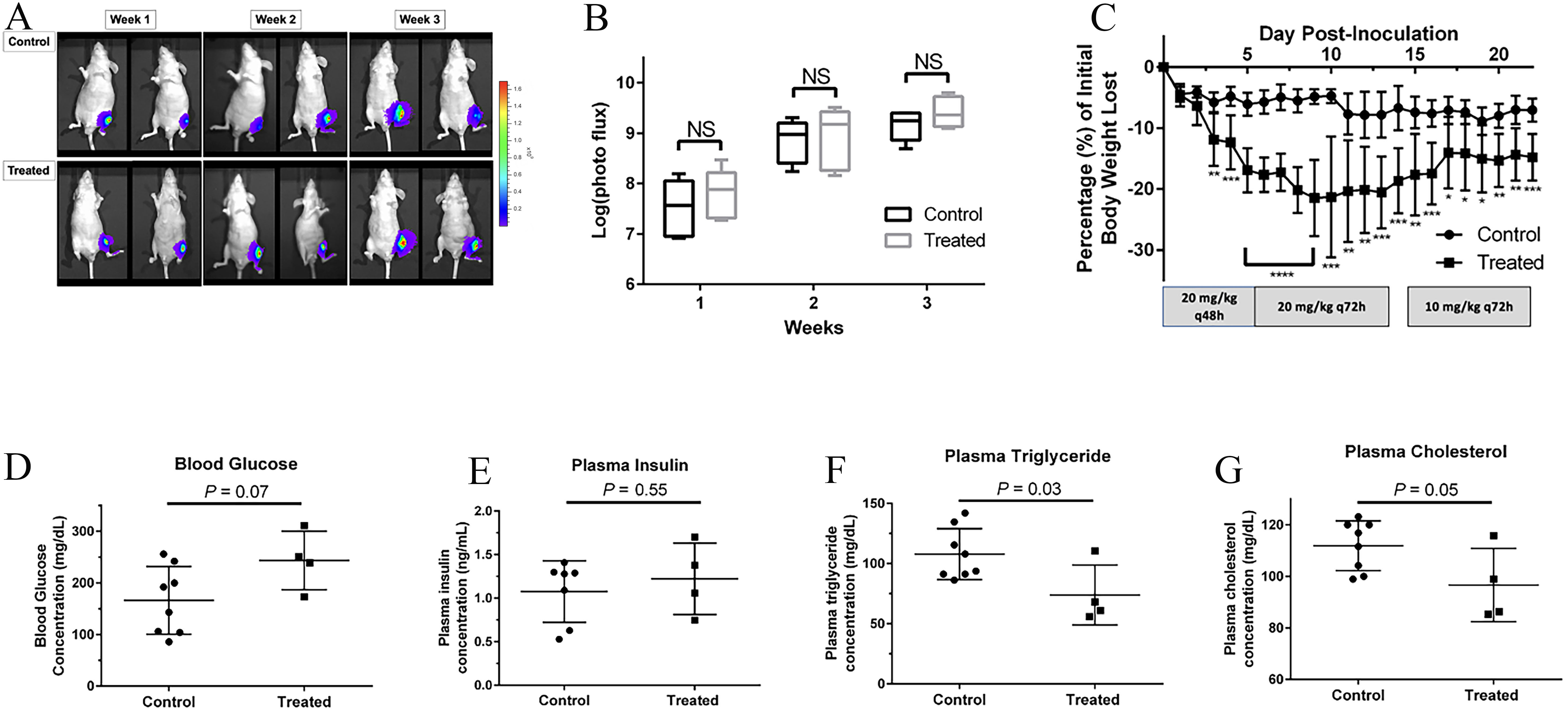
Figure 3. In vivo efficacy and safety of DRB18. Ace-1YFP-LUC cells were injected into the proximal tibias of male, 8-week-old nude mice (n = 16) initially treated with 20 mg/kg DRB18 or DMSO/PBS (1:1; v:v) for three weeks by intraperitoneal injection every 48 h. Surviving mice (n = 12) were sacrificed after 3 weeks. (A) Representative bioluminescent images of control (top row) and treated (bottom row) mice. Images were taken once weekly for 3 weeks. Bioluminescent intensity is proportional to the number of viable tumor cells and reflects tumor size. The photon flux (photons/second/region of interest) scale bar applies only to the left adjacent images; (B) Quantification of bioluminescent intensity for control and treated mice for each week of the study. DRB18 did not reduce the growth of intratibial tumors compared to controls. Box plot. NS: Not significant. Unpaired t-test at each time point; (C) Change in body weight between control and treated mice from study start to termination. The DRB18 concentration administered and frequency are listed below. q: quaque. Unpaired t-tests; (D-G) Changes in treatment-related metabolic biochemical parameters at the study’s termination between control (n = 8) and treated (n = 4) mice. Unpaired t-tests. *P < 0.05; **P < 0.01; ***P < 0.001; ****P < 0.0001.


