fig1

Progression of bone-metastatic prostate cancer in a mouse model treated with a novel pan-class I GLUT inhibitor (DRB18)

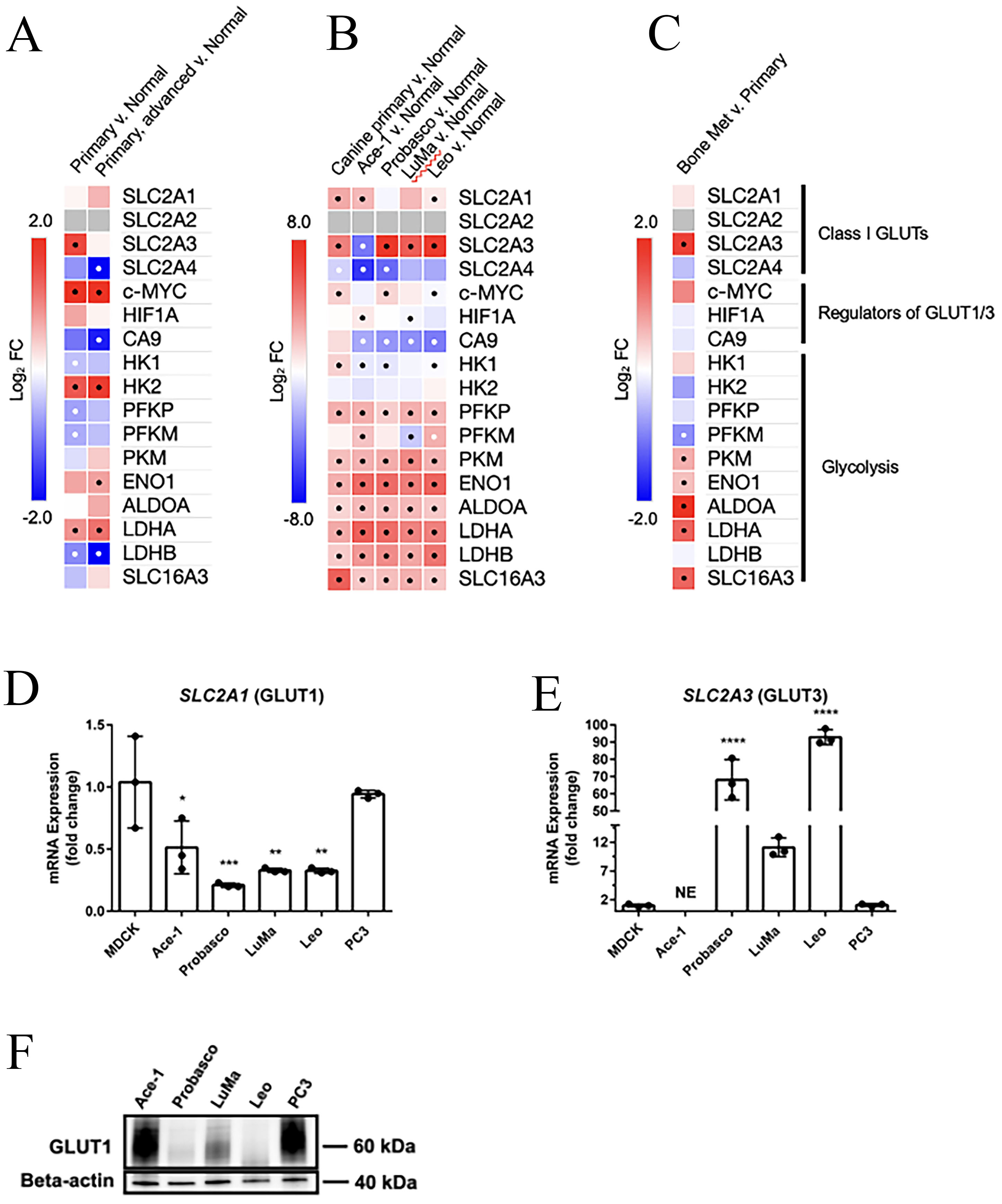
Figure 1. Expression of class I GLUTs and associated pathways in human and canine PCa and cell lines. (A-C) Gene expression heatmaps were generated from publicly available human or canine clinical samples and canine PCa cell lines. “Normal” indicates non-cancerous prostate gland for the respective species, human or canine. Scale bars indicate the log2 fold change (Log2 FC) in gene expression between groups, as displayed above the heatmaps. Black or white dots indicate the significance of P < 0.05 or smaller reached between groups; Gray boxes indicate the gene was not detected. (D) and (E) Gene expression of GLUTs 1 and 3 was confirmed by qRT-PCR of human and canine PCa cell lines in vitro. Cells were seeded in triplicate and normalized to the housekeeping gene, GAPDH. The relative expression of each cell line was calculated and statistically analyzed by one-way ANOVA relative to control (MDCK) cells. Data were displayed as mean ± SD. *P < 0.05; **P < 0.01; *** P < 0.001; ****P < 0.0001. NE: not expressed. (F). GLUT1 was produced by PCa cell lines in vitro. GLUTs: Glucose transporters; PCa: Prostate cancer; MDCK: Madin-Darby canine kidney cells.

Journal of Cancer Metastasis and Treatment
ISSN 2454-2857 (Online) 2394-4722 (Print)

Portico

All published articles are preserved here permanently:

https://www.portico.org/publishers/oae/

Portico

All published articles are preserved here permanently:

https://www.portico.org/publishers/oae/