


fig20
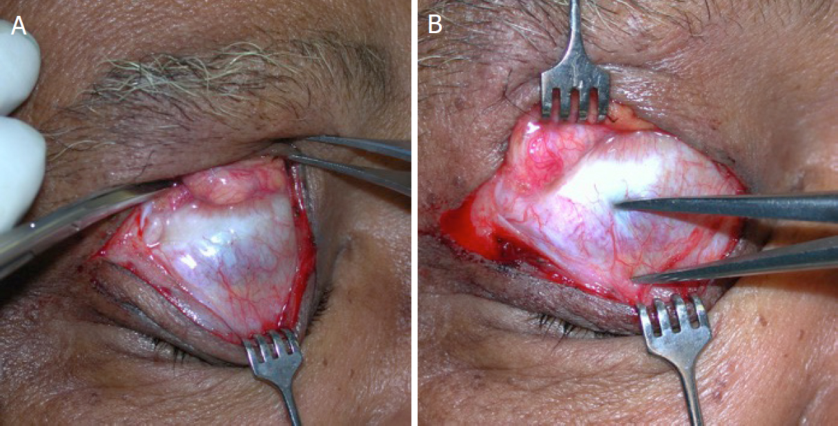
Figure 20. A: the dystrophic, thin levator is demonstrated; B: the calipers are used to show 10 mm of dehiscence from the tarsal plate to the levator aponeurosis
Figure 20. A: the dystrophic, thin levator is demonstrated; B: the calipers are used to show 10 mm of dehiscence from the tarsal plate to the levator aponeurosis


All published articles are preserved here permanently:
https://www.portico.org/publishers/oae/

